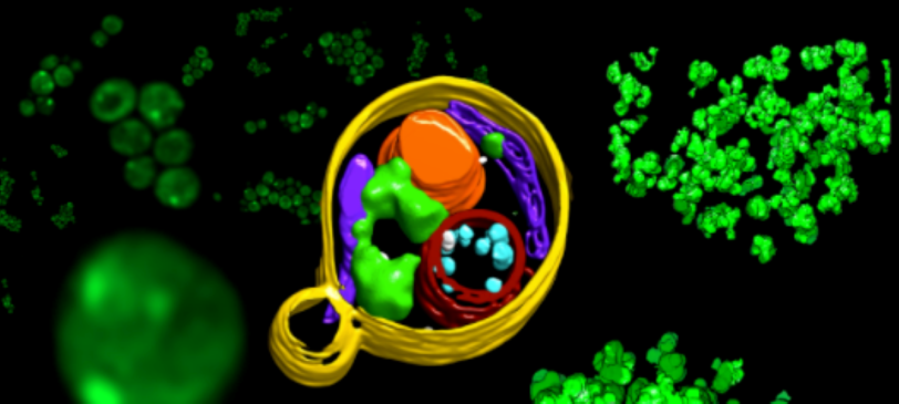
Cryogenic Electron Tomography (cryoET) is an emerging technique that can resolve subcellular structures in situ with the potential to reach near atomic resolutions. Correlated cryo-fluorescence light microscopy (cryoFLM) and cryoET of frozen, hydrated cells can be used to study cellular and molecular functions and dynamics in the 3D context of cells and tissues at a higher resolution than any other imaging technique. For samples which are too thick for imaging via cryoET, a focused ion beam (FIB) can be used to produce thin lamellae (100-300 nm) from vitrified cells or tissues containing targets of interest. The embedded cryoFLM in the cryoFIB can guide the search of the targets to form the lamellae which are then imaged by cryoET in a transmission electron microscope.
MISSION STATEMENT:
The mission of the Stanford-SLAC CryoET Specimen Preparation Center (SCSC) is to provide:
- Access to state-of-the-art instrumentation and knowledge about cryoET specimen preparation
- Training and support, to increase the ability of the cryoET community to perform experiments particularly in Life and Biomedical Sciences
- Development of workflows and implementation of new technologies for cryoET specimen preparation
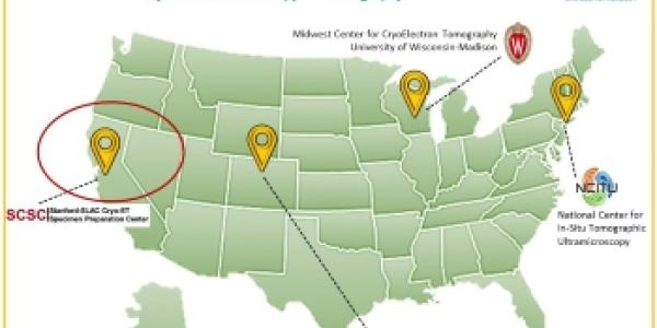
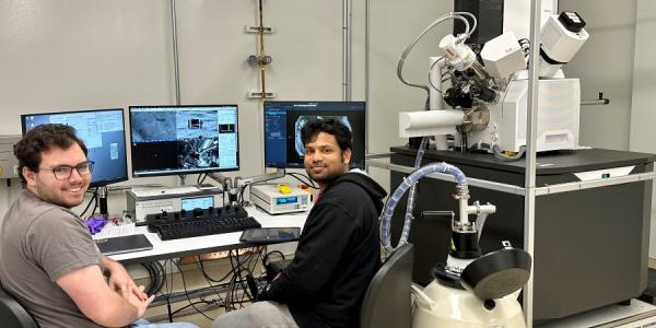
The Center implements state-of-the-art cryo-specimen preparation devices. We provide expert staff that train, assist, and advise users both onsite and remotely. We cross-train scientists who want to employ cryoET within their own research portfolios. Our training is targeted at a wide variety of skill levels - including short to long-term in-residence training programs for cryoET specimen preparation. Access to the Center is through an open process based on scientific merit. Specimens prepared at SCSC can be transferred for data collection at the investigator's lab or at the Center Hub in Madison, Wisconsin.
About
SCSC is one of the four service centers of a National Network for Cryoelectron Tomography...

Facilities and Resources
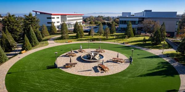
SCSC is located on the beautiful SLAC campus of Stanford University. The equipment, laboratories...

Access
Application to SCSC is through a unified application process for all four of the sevice centers...

SCSC NIH Support Acknowledgement
The SCSC is supported by the National Institutes of Health Common Fund Transformative High Resolution Cryo-Electron Microscopy and Cryo-Electron Tomography program through Grant number U24GM139166.
