
Facilities and Resources
SCSC CryoET Facilities
Equipment*
CryoEM specimen preparation devices, cryo-fluorescence optical microscopes, cryo-focused ion beam scanning electron microscopes, and 200/300 kV electron cryo-microscopes equipped with direct electron detectors, energy filters, and phase plates. (see photos of available instruments). SCSC users will have access to these instruments after proper training by our SCSC staff.
- High Pressure and Plunge Freezing Instruments including Leica EM ICE, Leica EM GP2, Thermo Fisher Vitrobot, and Gatan CP3 plunger in a low humidity laboratory.
- Cryo Fluorescent Light Microscopy (cryoFLM) includes a Leica Stellaris 8 microscope for high resolution cryoFLM imaging. Additionally, a Delmic METEOR integrated FLM is available on the Aquilos 2 cryoFIB-SEM, and a ThermoFisher iFLM on the Hydra Bio PFIB-SEM.
- Cryo Focused Ion Beam Scanning Electron Microscopes (CryoFIB-SEM) include a ThermoFisher Aquilos 2, and a Hydra Bio Plasma FIB-SEM (PFIB), both with integrated Fluorescence Light Microscopes (FLM). A Delmic METEOR FLM is integrated with the Aquilos2, and a ThermoFisher iFLM with the Hydra Bio PFIB-SEM.
- A Leica VT1200S vibratome is available for slicing of tissue samples before high pressure freezing.
- Cryo-Sectioning Ultramicrotome Leica UC6 equipped with a cryo chamber.
- CryoEM Titan Krios Galcios 2 electron microscopes are equipped with Volta phase plate, energy filter (BioQuantum or Selectris), and direct electron detector (K3 or Falcon 4i camera). ✅
*Full list of SCSC Instrumentation
Biological Laboratory Facilities:
SCSC has a tissue culture laboratory, cold room and refrigeration facilities, and standard biochemistry instrumentation available for onsite users. These facilities are BSL-2 compliant.
Data Management and Computational Resources:
All experimental data collected under SCSC will be held on disk for two months from the day of collection. The data is transferred to the data cluster as soon as it is collected and is therefore immediately available for download via either the usual Unix tools (RSYNC, SCP, BBCP, etc.) or through Globus. More details regarding data transfer can be found at https://confluence.slac.stanford.edu/x/mYoYDg.
After two months, the data is transferred onto tape and will be kept for 22 additional months. Requests for data retrieval after the data is moved onto tape can be made via email to unix-admin@slac.stanford.edu with specific information regarding the date of the data collection, the proposal number, and the microscope that was used.
Two years after the data has been collected, the data will be purged completely from our systems.

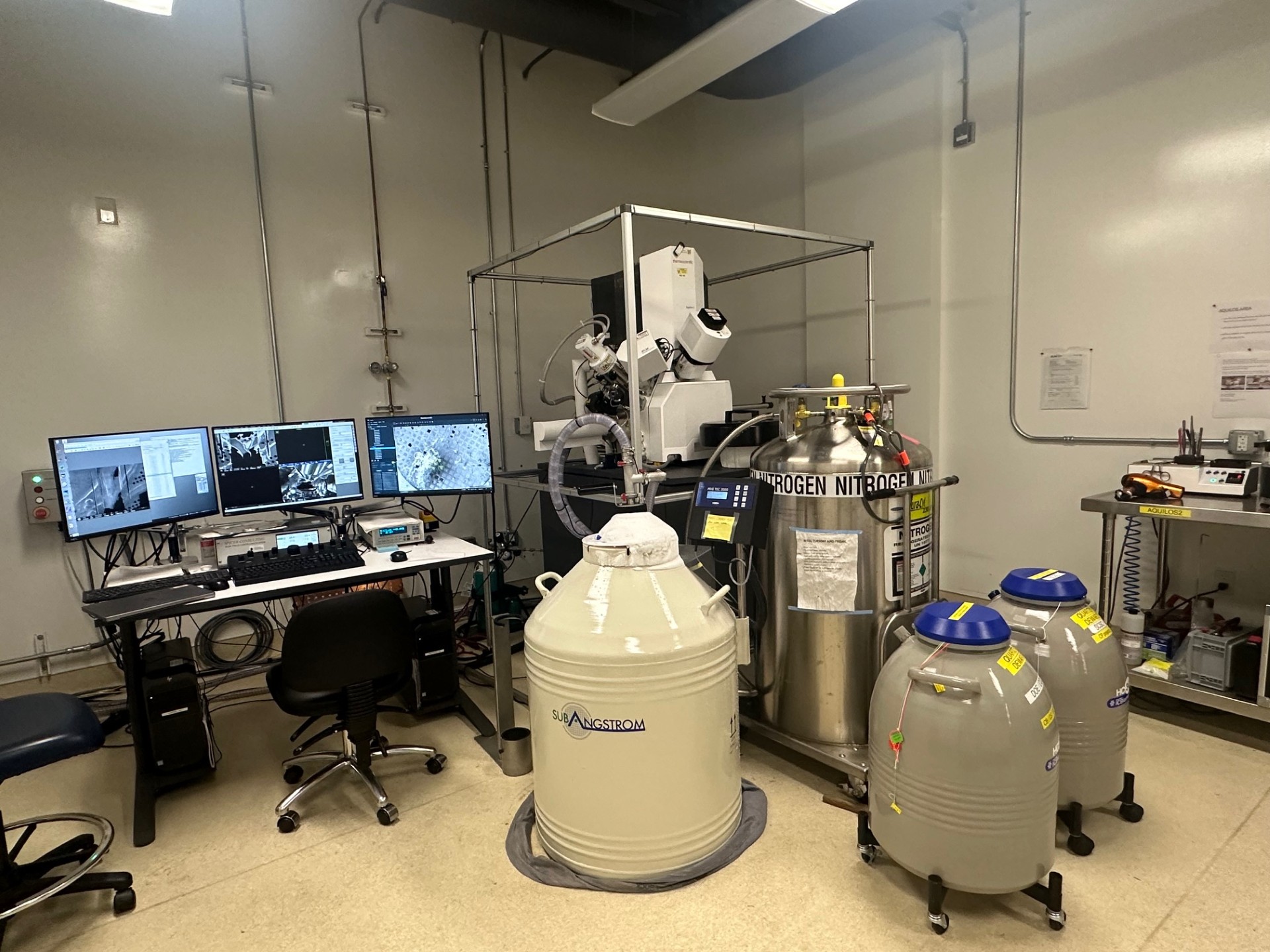
Aquilos 2 FIB2

The Aquilos 2 cryo-FIB is housed alongside the Hydra Bio P-FIB in a humidity-controlled room in Bldg. 006 and is certified to work with BSL2 samples. Both the Aquilos 2 and Hydra have embedded cryo-fluorescence light microscopes (cryo-FLM) - The cryo-FLM in Aquilos 2 is a METEOR from Delmic, while Hydra Bio P-FIB is an iFLM from Thermo Fisher. The cryo-FIBs are connected to a SubAngstrom GEN2 dewar for extended and multi-day operation.
photo taken by Dr. Lydia Joubert

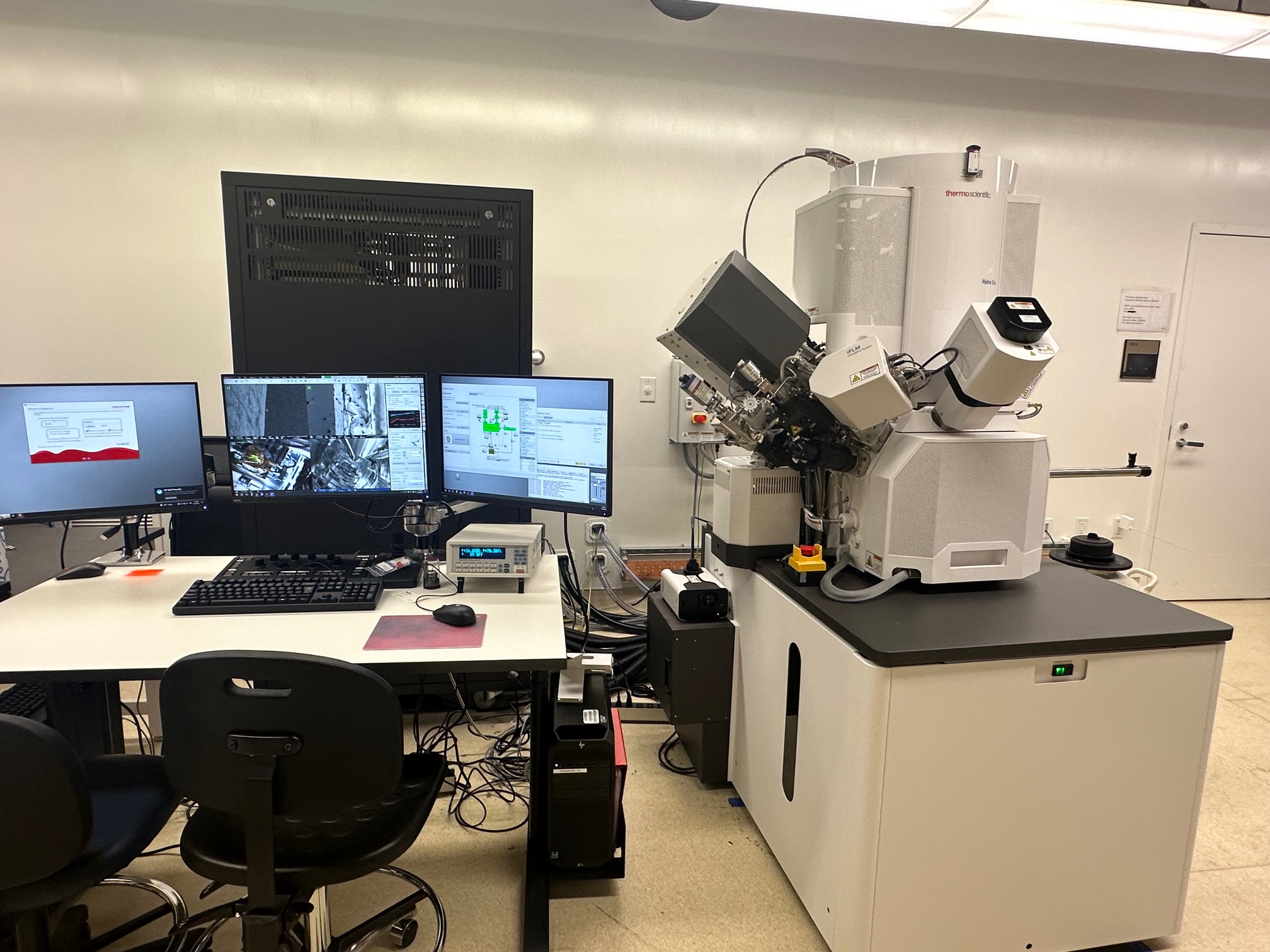
Hydra Bio P-FIB

The Hydra Bio P-FIB is housed alongside the Aquilos 2 in a humidity-controlled room in Bldg. 006 and is certified to work with BSL2 samples. Both the Hydra and Aquilos 2 have embedded cryo-fluorescence light microscopes (cryo-FLM) - The cryo-FLM in Aquilos 2 is a METEOR from Delmic, while Hydra Bio P-FIB is an iFLM from Thermo Fisher. The cryo-FIBs are connected to a SubAngstrom GEN2 dewar for extended and multi-day operation.
photo taken by Dr. Alexandre Cassago

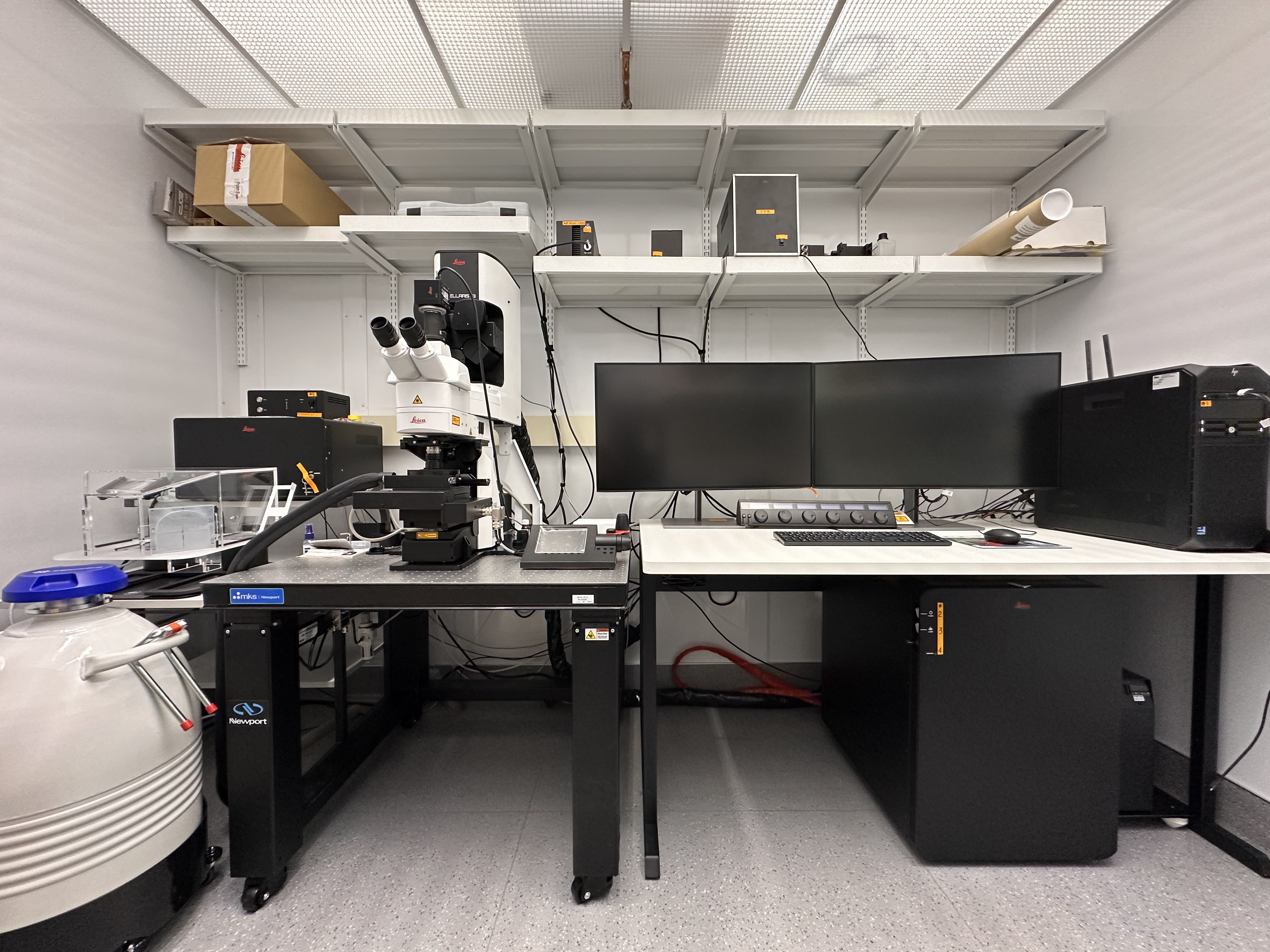
Leica Stellaris Cryo-confocal system

The Leica STELLARIS Cryo-confocal system was our recent acquisition to enhance the quality and resolution of the confocal images of fluorescence labelled samples which have been vitrified prior to loading into the cryoFIB-SEM. The Stellaris is housed in a humidity-controlled room in Bldg. 006 and is certified to work with BSL2 samples.
photo taken by Dr. Alexandre Cassago

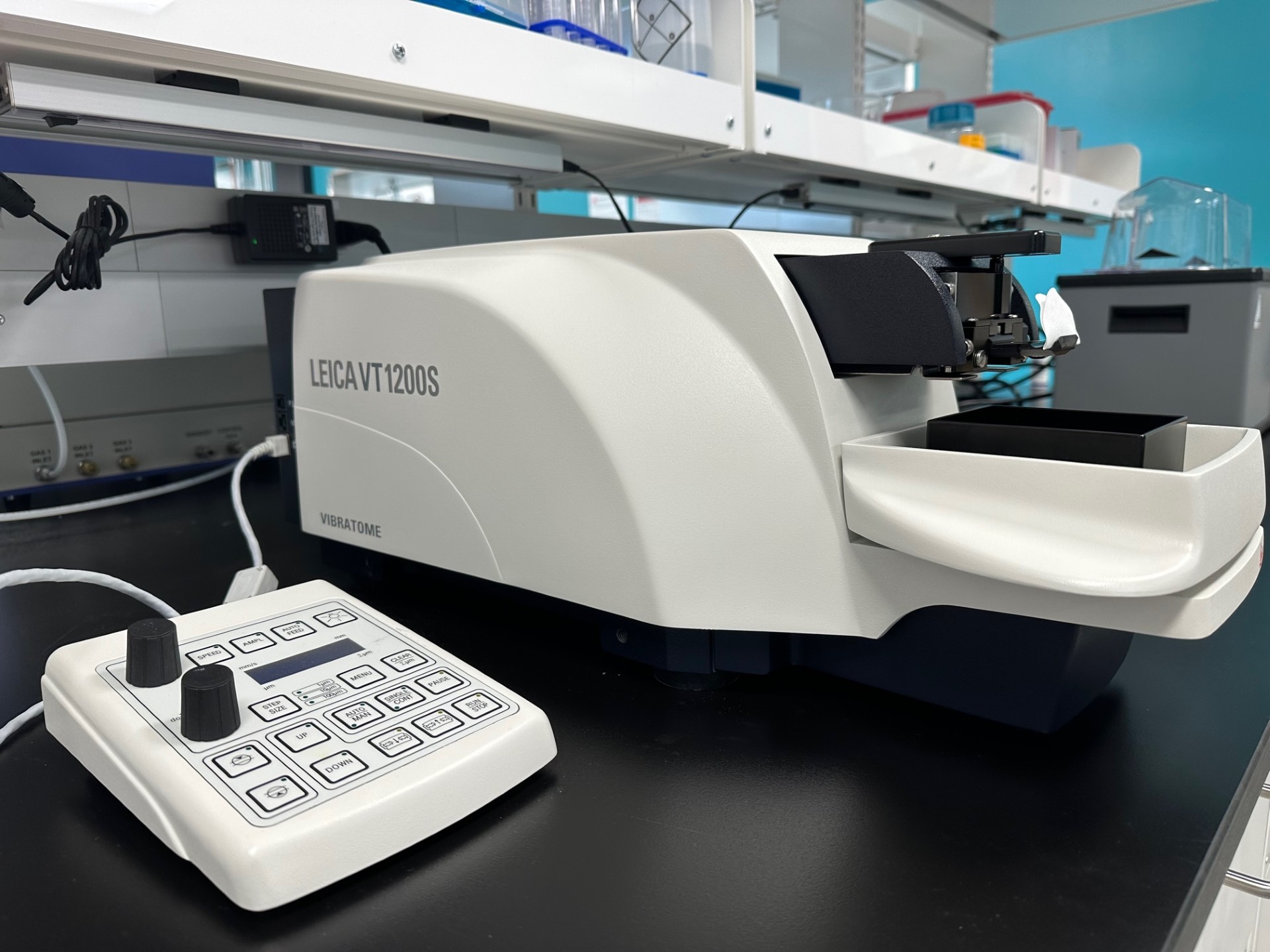
Leica VT1200S vibratome.jpg

A Leica VT1200S vibratome is available for slicing of tissue samples before high pressure freezing.
photo taken by Dr. Alexandre Cassago

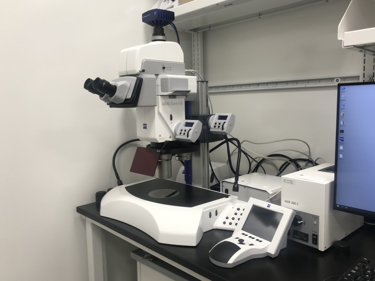
Zeiss AxioZoom.jpg

Brightfield and Fluorescence dissection microscope for evaluating cell cultures before vitrification: Axio Zoom.V16 gives superior fluorescence brightness in large object fields. With Plan-NEOFLUAR Z 2.3x you achieve a numerical aperture of NA 0.5 in an object field of 1.5 millimeters. One can get superior brightness in large object fields and view complete model organisms in fluorescence contrast.
photo taken by Dr. Alexandre Cassago

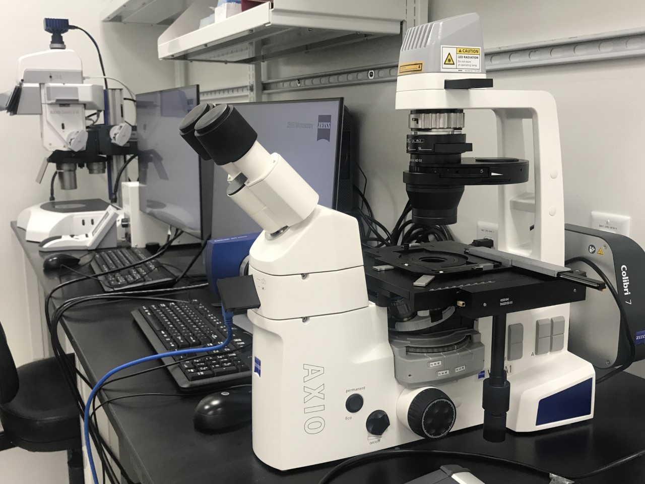
Zeiss AxioVert.jpg

Inverted Brightfield and Fluorescence microscope for evaluating cell cultures before vitrification: The AxioVert is an inverted light microscope with all standard contrasting techniques, including DIC, to investigate cell cultures.
photo taken by Dr. Alexandre Cassago

Leica_ACE600_Sputter_Coater_001.jpg

Sputter-coating device for grid preparation, including Au, Pt, Pd and Ir targets, as well as C thread.
Operated at room temperature, the sputter-coater is a versatile high vacuum film deposition instrument used to produce nanometer thin but robust carbon films, deposit fine-grained metal layers and perform glow discharging. At our center it is mainly used for grid preparation in the waffle milling workflow.
photo taken by Dr. Alexandre Cassago

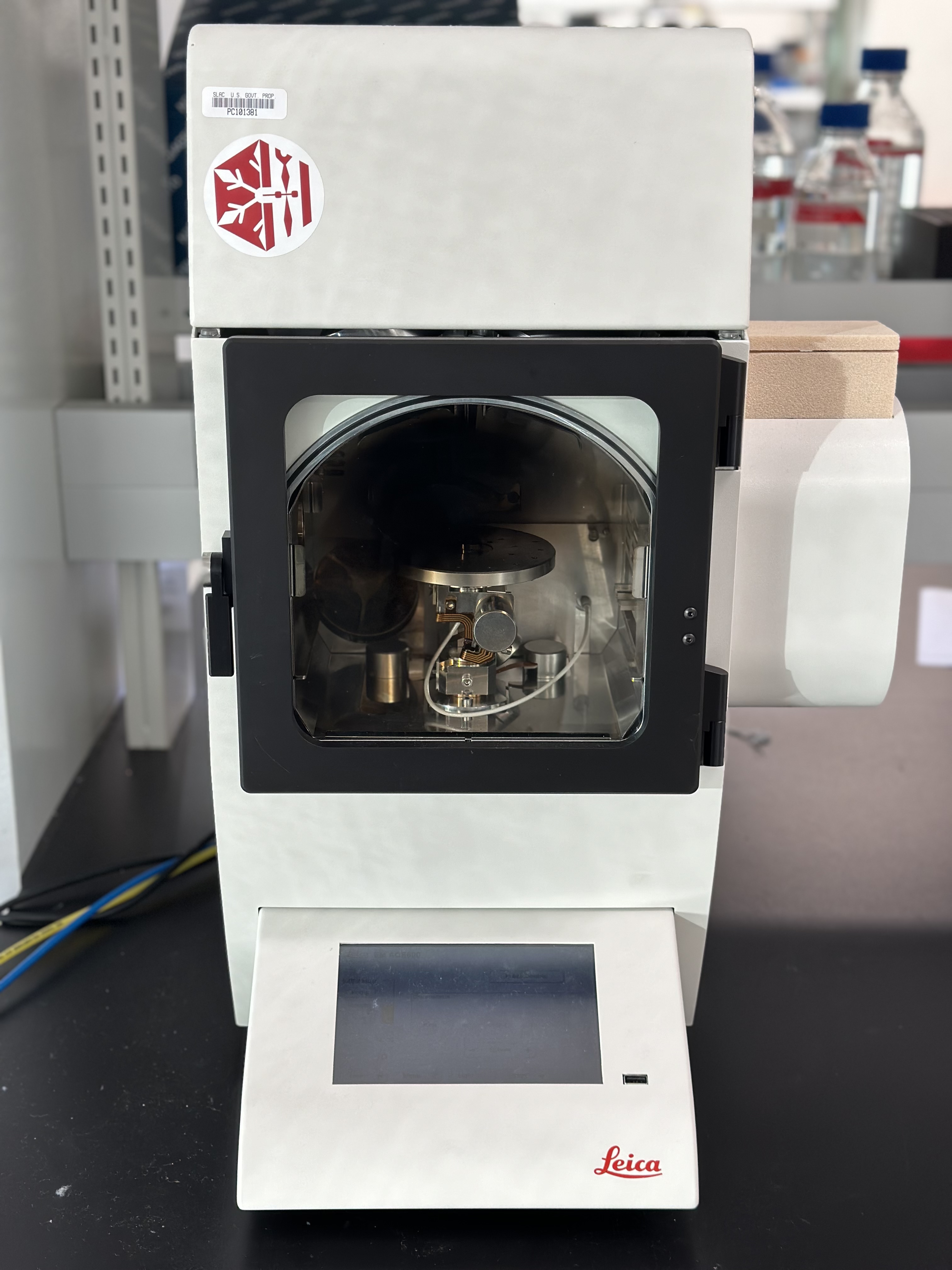
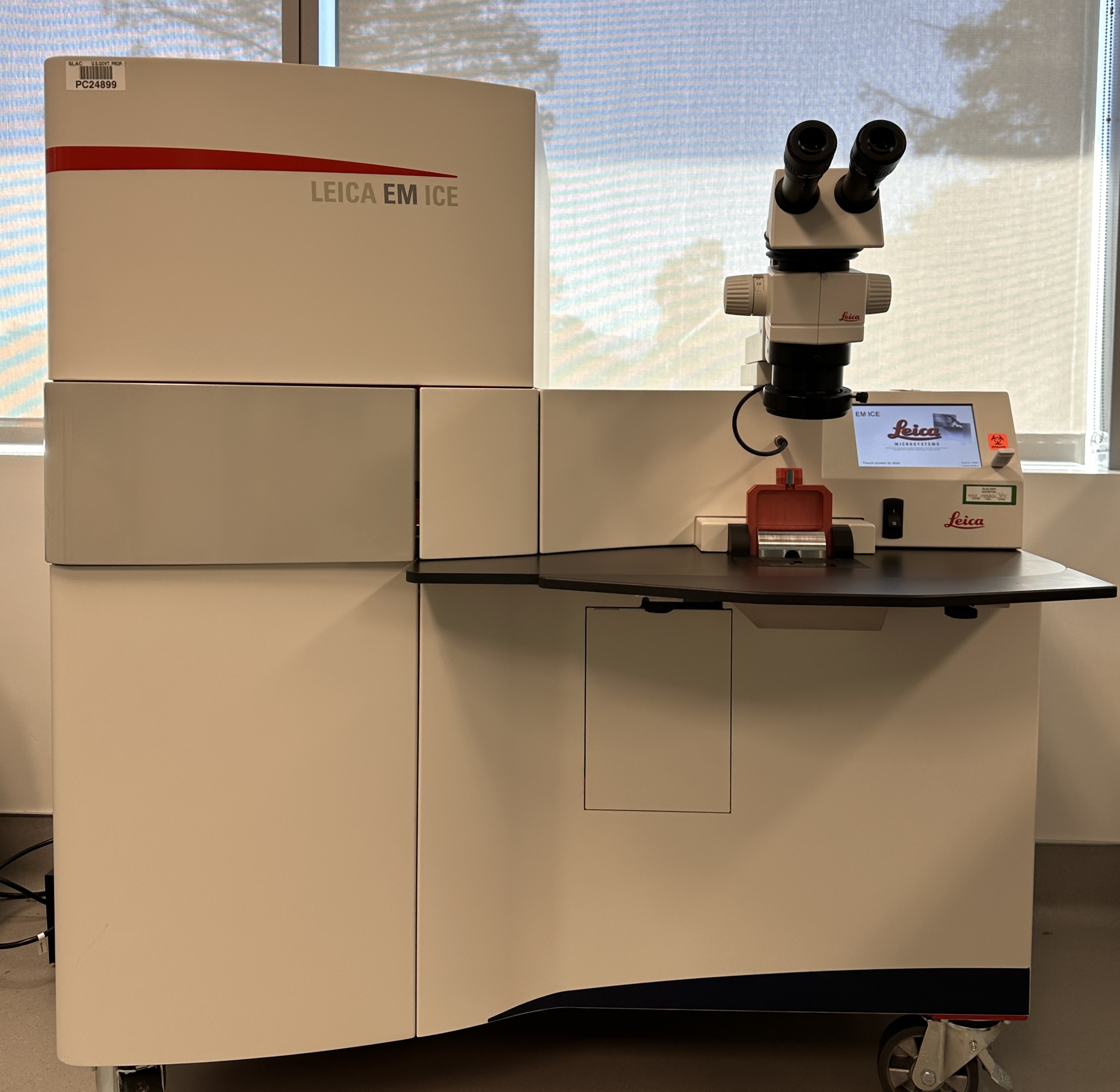
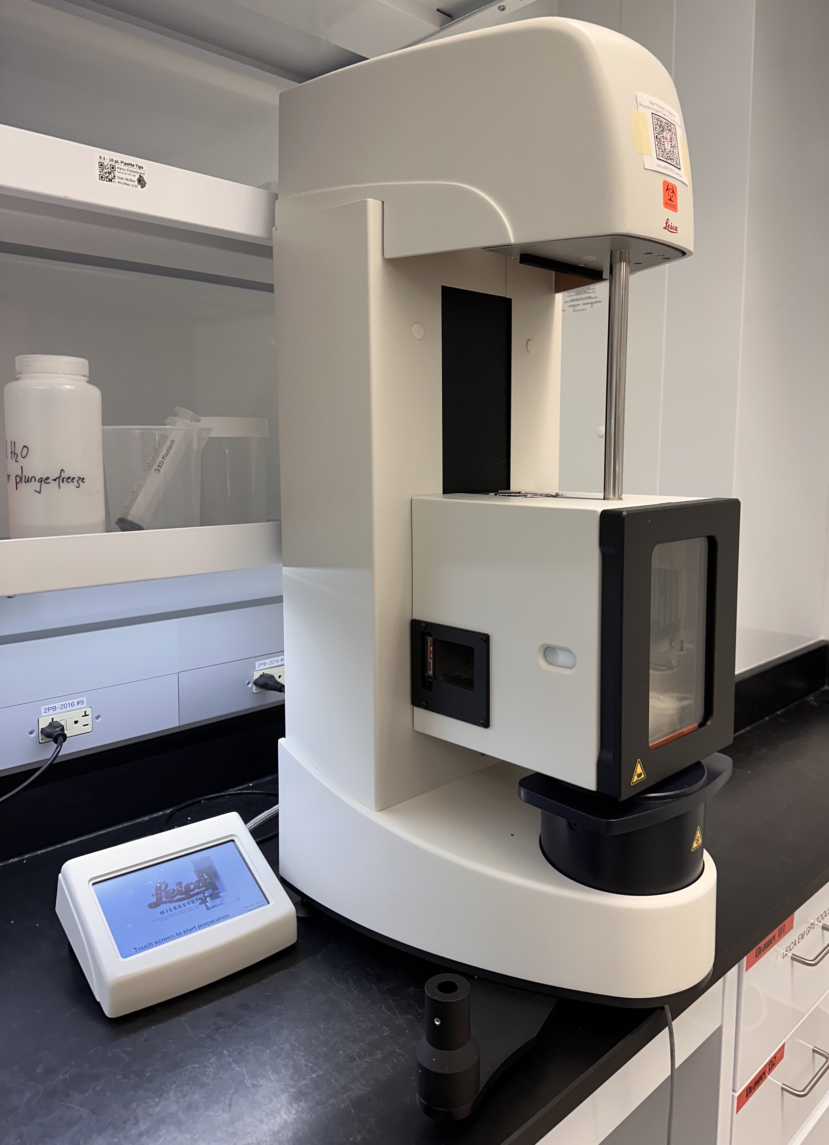
High_Pressure_Freezer_003.jpg

High pressure freezing and waffle preparation of specimens:
The Leica EM ICE provides superior and repeatable high pressure freezing of samples up to 200microns thick and 5mm in diameter, in an ergonomically designed workstation combined with a stereomicroscope for ease of loading and manipulating of samples.
The Leica ICE HP Freezer has become an essential component in our cryo ‘Mill and View’ workflow development for tissue and organoid specimens, applicable to health and disease models.
photo taken by Dr. Alexandre Cassago

